
Page 68 - 22-0424
P. 68
The International Journal of the Royal Society of Thailand
Volume XI - 2019
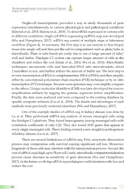
Single-cell transcriptomic provided a way to study thousands of gene
expression simultaneously in various physiological and pathological conditions
(Kiryluk et al., 2018; Malone et al., 2018). To detect RNA expression in various cells
in different conditions, single-cell RNA sequencing (scRNA-seq) was developed
(Wu and Humphreys, 2017). scRNA-seq consist of multiple steps in laboratory
workflow (Figure 4). In summary, the first step is to use enzyme to lyse biopsy
tissue into single cell and then put the cell in compartment such as plate/tube or
microfluidic. Plate or tube based are costly due to use of large amount of tube/
well and buffer. Fluidigm C1 system can capture larger amount of cells in the
chambers and reduce the cost (Islam et al., 2014; Wu et al., 2014). Microfluidic
techniques incorporate cells and barcoded DNA into microdroplets, about
2 nanoliters in size, and further reduce the amount of buffer use. The next step is
reverse transcription of RNA to complemaentary DNA (cDNA) and then amplify
either by conventional polymerase chain reaction (PCR) technique or by in vitro
transcription (IVT) technique. Because some genomes may over amplify compare
to the others. Unique molecular identifiers (UMI) was later developed for remove
amplification artifacts by tagging the genome segments before amplification.
Finally, the data were analyzed and were compared to the existing database by
specific computer software (Liu et al., 2014). The details and advantages of each
methods were previously reviewed elsewhere (Wu and Humphreys, 2017).
One of the example studies of scRNA-seq in kidney disease was done by
Lu et al. They performed snRNA-seq analysis of mouse mesangial cells using
the fluidigm C1 platform. They found heterogeneity among mesangial cells with
correlation coefficients of only 0.20. They also found 1045 genes that appear in
every single mesangial cells. These finding created a new insight in pathogenesis
of kidney disease (Lu et al., 2017).
There are several limitations of scRNA-seq. First, enzymatic dissociation
process may compromise cells survival causing significant cell loss. Moreover,
fragments of these cells may interfere with the interpretation process. Second, the
cost of scRNA-seq is high, up to 5$ per cell. Lastly, microfluidic compartmentalization
process cause decrease in sensitivity of gene detection (Wu and Humphreys,
2017). In the future, evolving scRNA-seq techniques could minimize cells loss and
reduce the cost.
62 Precision Medicine in Acute kidney injury
11/7/2565 BE 13:28
_22-0424(055-076)7.indd 62 11/7/2565 BE 13:28
_22-0424(055-076)7.indd 62