
Page 79 - The lraternational Journal of the Royal Society of Thailand.indd
P. 79
The International Journal of the Royal Society of Thailand
Volume XV-2023
The first example is metastatic carcinoma with squamous differentiation
mimicking PBL. The tumor cells are positive for CD138 and questionable restriction to
cytoplasmic immunoglobulin kappa light chain. However, upon the review, it is
non-specific cytoplasmic staining for both kappa and lambda light chains as the
reactive plasma cells should not have tumor cell morphology. The tumor cells are in
fact positive for AE1/AE3 and p40 protein, indicative of squamous differentiation
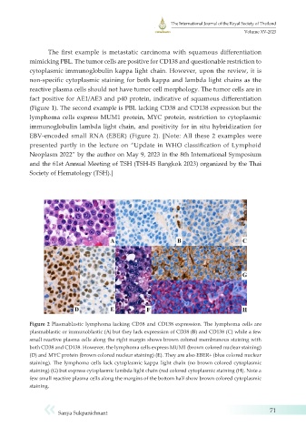
(Figure 1). The second example is PBL lacking CD38 and CD138 expression but the
lymphoma cells express MUM1 protein, MYC protein, restriction to cytoplasmic
immunoglobulin lambda light chain, and positivity for in situ hybridization for
EBV-encoded small RNA (EBER) (Figure 2). [Note: All these 2 examples were
presented partly in the lecture on “Update in WHO classification of Lymphoid
Neoplasm 2022” by the author on May 9, 2023 in the 8th International Symposium
and the 61st Annual Meeting of TSH (TSH-IS Bangkok 2023) organized by the Thai
Society of Hematology (TSH).]
Figure 2 Plasmablastic lymphoma lacking CD38 and CD138 expression. The lymphoma cells are
plasmablastic or immunoblastic (A) but they lack expression of CD38 (B) and CD138 (C) while a few
small reactive plasma cells along the right margin shows brown colored membranous staining with
both CD38 and CD138. However, the lymphoma cells express MUM1 (brown colored nuclear staining)
(D) and MYC protein (brown colored nuclear staining) (E). They are also EBER+ (blue colored nuclear
staining). The lymphoma cells lack cytoplasmic kappa light chain (no brown colored cytoplasmic
staining) (G) but express cytoplasmic lambda light chain (red colored cytoplasmic staining (H). Note a
few small reactive plasma cells along the margins of the bottom half show brown colored cytoplasmic
staining.
Sanya Sukpanichnant 71