
Page 78 - The lraternational Journal of the Royal Society of Thailand.indd
P. 78
The International Journal of the Royal Society of Thailand
Volume XV-2023
MUM1, CD138, BLIMP1), and 3) negativity for CD20, PAX5, ALK and KSHV/HHV8
(WHO Classification of Tumours Editorial Board, 2022). If immunostaining for
KSHV/HHV8 is not available then it cannot completely exclude KSHV/HHV8+ DLBCL
before making a diagnosis of PBL. Importantly, the first essential diagnostic criterion
for PBL needs to mark sure that it is lymphoma because it is well documented that
plasmablast or immunoblast may be difficult to distinguish from large tumor cells in
other non-hematologic malignancies such as carcinoma, melanoma, or sarcoma.
Moreover, melanoma cells express MUM1 protein and both carcinoma and melanoma
cells express CD138. [Kind et al, 2019; Natkunam et al, 2001] We recently published a
case report of metastatic prostatic adenocarcinoma mimicking PBL as the tumor cells
resembled plasmablasts and they were positive for CD138 but negative for CD3, CD20,
CD30, CD45, CK7, and CK20 so that the first pathologist gave the diagnosis of PBL.
But we found that the tumor cells lack cytoplasmic immunoglobulin light chains,
either of kappa or lambda. In addition, the tumor cells were positive for AE1/AE3,
CK19, PSA and NKX3.1, indicative of metastatic prostatic adenocarcinoma.
[Sukpanichnant et al, 2022] Figures 1 and 2 are two more examples to illustrate
problems to diagnose PBL in daily practice.
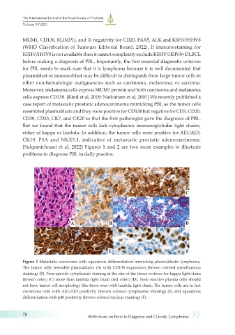
Figure 1 Metastatic carcinoma with squamous differentiation mimicking plasmablastic lymphoma.
The tumor cells resemble plasmablasts (A) with CD138 expression (brown colored membranous
staining) (B). Non-specific cytoplasmic staining at the rim of the tissue sections for kappa light chain
(brown color) (C) more than lambda light chain (red color) (D). Note reactive plasma cells should
not have tumor cell morphology like those seen with lambda light chain. The tumor cells are in fact
carcinoma cells with AE1/AE3 positivity (brown colored cytoplasmic staining) (E) and squamous
differentiation with p40 positivity (brown colored nuclear staining) (F).
70 Reflections on How to Diagnose and Classify Lymphoma